
Understanding Fetal Monitor Probe Types and Core Clinical Use Cases
Doppler, Fetoscope, and Internal Probes: When Each Is Indicated in Prenatal and Intrapartum Settings
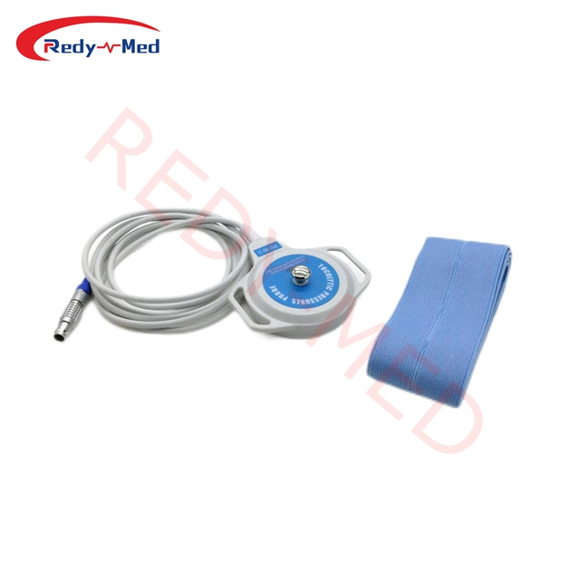
Fetal monitoring relies on three primary probe types—Doppler ultrasound, fetoscope, and internal probes—each matched to distinct clinical needs. Doppler probes are the standard for routine prenatal visits and early labor due to their portability, ease of use, and non-invasive operation. Fetoscopes—acoustic stethoscopes requiring no power or gel—support intermittent auscultation in low-risk pregnancies, especially where minimal technology use aligns with care philosophy or resource constraints. Internal probes, such as fetal scalp electrodes (FSE), are reserved for active labor when continuous, high-fidelity data is essential and external monitoring is unreliable—common with high maternal BMI, excessive fetal movement, or indeterminate heart rate patterns. Placement requires ruptured membranes and carries a small but documented increase in infection risk compared to external methods. As outlined in ACOG Practice Bulletin No. 189 and NICE Guideline NG123, internal monitoring offers superior accuracy in detecting subtle signs of fetal compromise during high-risk deliveries—but only when clinically justified.
Frequency Selection (2 MHz, 3 MHz, 5 MHz): Aligning Fetal Monitor Probe Specs with Gestational Age and Maternal Anatomy
Ultrasound frequency selection directly affects signal penetration and resolution—and must be tailored to gestational age and maternal anatomy. A 2 MHz probe provides deeper tissue penetration, making it optimal for early pregnancy (<20 weeks) or patients with BMI ≥30 kg/m², where adipose tissue attenuates higher-frequency signals. The 3 MHz probe strikes a practical balance between depth and clarity for mid-pregnancy (20–30 weeks) in average-weight patients. At 5 MHz, resolution improves significantly, ideal for late gestation (>30 weeks) when the fetus is closer to the abdominal wall—particularly in lean patients. Using mismatched frequencies introduces artifacts: for example, applying 5 MHz in obesity often yields weak or absent signals, while 2 MHz in late-term, low-BMI patients may blur fine waveform details. Clinicians should reassess frequency choice at each trimester transition and whenever maternal weight or fetal position changes substantially.
Optimizing Fetal Monitor Probe Performance Through Proper Placement and Signal Management
Best Practices for Belt Positioning, Acoustic Coupling, and Patient Positioning to Maximize FHR Detection
Accurate FHR detection depends on three interdependent technical factors: transducer placement, acoustic coupling, and patient positioning. Begin by locating the fetal back via Leopold’s maneuvers—then place the probe just below the maternal umbilicus, adjusting laterally or vertically based on fetal lie and station. Secure the belt snugly enough to prevent slippage but loose enough to allow natural respiratory motion; overtightening induces pressure artifacts and discomfort. Apply generous, uniform ultrasound gel to eliminate air pockets—reapply as needed if signal quality declines. For optimal uterine perfusion and fetal mobility during non-stress testing, position the patient in left lateral tilt (15–30°). In obese patients, combine semi-Fowler’s positioning with gentle hip flexion to reduce abdominal wall tension and improve probe-skin contact.
Common Signal Artifacts—Maternal BMI, Fetal Position, and Amniotic Fluid Volume—and How to Mitigate Them
Signal degradation most commonly stems from maternal BMI >30 kg/m², occiput posterior fetal position, or oligohydramnios (AFI <5 cm). High BMI causes significant ultrasound attenuation—counteract this by selecting a 2 MHz probe, incrementally increasing transducer pressure, and repositioning to anatomical “windows” (e.g., flank or lower abdomen). For posterior presentations, encourage hands-and-knees positioning for 10–15 minutes to promote spontaneous rotation; reassess after. With low amniotic fluid volume (<200 mL), elevate the maternal pelvis using a wedge to centralize fetal parts near the probe surface. Motion artifacts from respiration or fetal activity respond best to real-time gain adjustment and built-in signal filtering—modern monitors flag inconsistent tracings automatically. If external tracing remains suboptimal after three structured repositioning attempts—including probe relocation, maternal position change, and gel reapplication—consider temporary internal monitoring per ACOG guidance.
Selecting a Fetal Monitor Probe Based on Clinical Workflow, Patient Experience, and Long-Term Value
Ambulatory Support, Waterproofing, and Ergonomic Design for Seamless Outpatient and Telehealth Prenatal Monitoring
Contemporary fetal monitor probes must support evolving models of care—including outpatient, home-based, and telehealth delivery. Ambulatory designs enable reliable FHR tracking during daily movement without compromising signal fidelity. Waterproofing allows safe use during bathing or showering, supporting longitudinal adherence in high-risk pregnancies where frequent monitoring is indicated. Ergonomic contours and low-profile sensors minimize skin irritation and improve overnight wearability—key drivers of sustained engagement. Research published in AJOG MFM (2023) found that 79% of patients with gestational hypertension or diabetes preferred wearable, ambulatory monitors over clinic-only devices, citing improved autonomy and reduced travel burden. For telehealth integration, prioritize probes with Bluetooth 5.0+ connectivity and automated, HIPAA-compliant data syncing to EHR-adjacent platforms—eliminating transcription errors and enabling timely clinician review. Battery life should exceed 24 hours to ensure uninterrupted overnight capture, and device compatibility with common smartphones or tablets ensures broad accessibility across diverse patient populations.
Cost–Utility Analysis: Reusability, Compatibility, and Total Ownership Cost Across OB/GYN and Midwifery Practice Models
Sustainable probe selection balances upfront cost, durability, interoperability, and lifecycle support. High-grade reusable probes deliver up to 93% cost savings over disposables within 18 months when sterilized according to FDA-cleared protocols (e.g., low-temperature hydrogen peroxide gas plasma). Cross-platform compatibility—especially Bluetooth 5.0+ and standardized output formats (e.g., HL7 or IEEE 11073)—prevents vendor lock-in and simplifies system upgrades. Total ownership costs vary meaningfully by practice model:
| Cost Factor | Midwifery Practice | Hospital OB/GYN |
|---|---|---|
| Initial Probe | $800–$1,200 | $1,500–$2,000 |
| Annual Maintenance | 8–12% of purchase | 5–8% of purchase |
| Training/Support | Low | High |
| Avg. Lifespan | 3–5 years | 2–3 years |
Smaller practices benefit most from modular, serviceable probes that avoid full-system replacement, while larger institutions should negotiate comprehensive service contracts covering firmware updates, remote diagnostics, and sterilization validation support. Critically, non-compliant sterilization has been linked to a 140% rise in unplanned probe replacements—making validation documentation a non-negotiable procurement criterion.
FAQ
Q: What are the main types of fetal monitor probes?
A: The main types are Doppler ultrasound, fetoscope, and internal probes, each suited to specific clinical scenarios.
Q: How do I select the appropriate ultrasound frequency for fetal monitoring?
A: Frequency choice depends on gestational age and maternal BMI, with 2 MHz for early pregnancy and high BMI, 3 MHz for mid-pregnancy, and 5 MHz for late gestation in lean patients.
Q: How can I optimize fetal monitor probe performance?
A: Optimal performance relies on proper transducer placement, adequate acoustic coupling with gel, and suitable patient positioning.
Q: What factors affect the cost and utility of fetal monitor probes?
A: Factors include initial cost, maintenance, training, durability, and compatibility with healthcare systems.